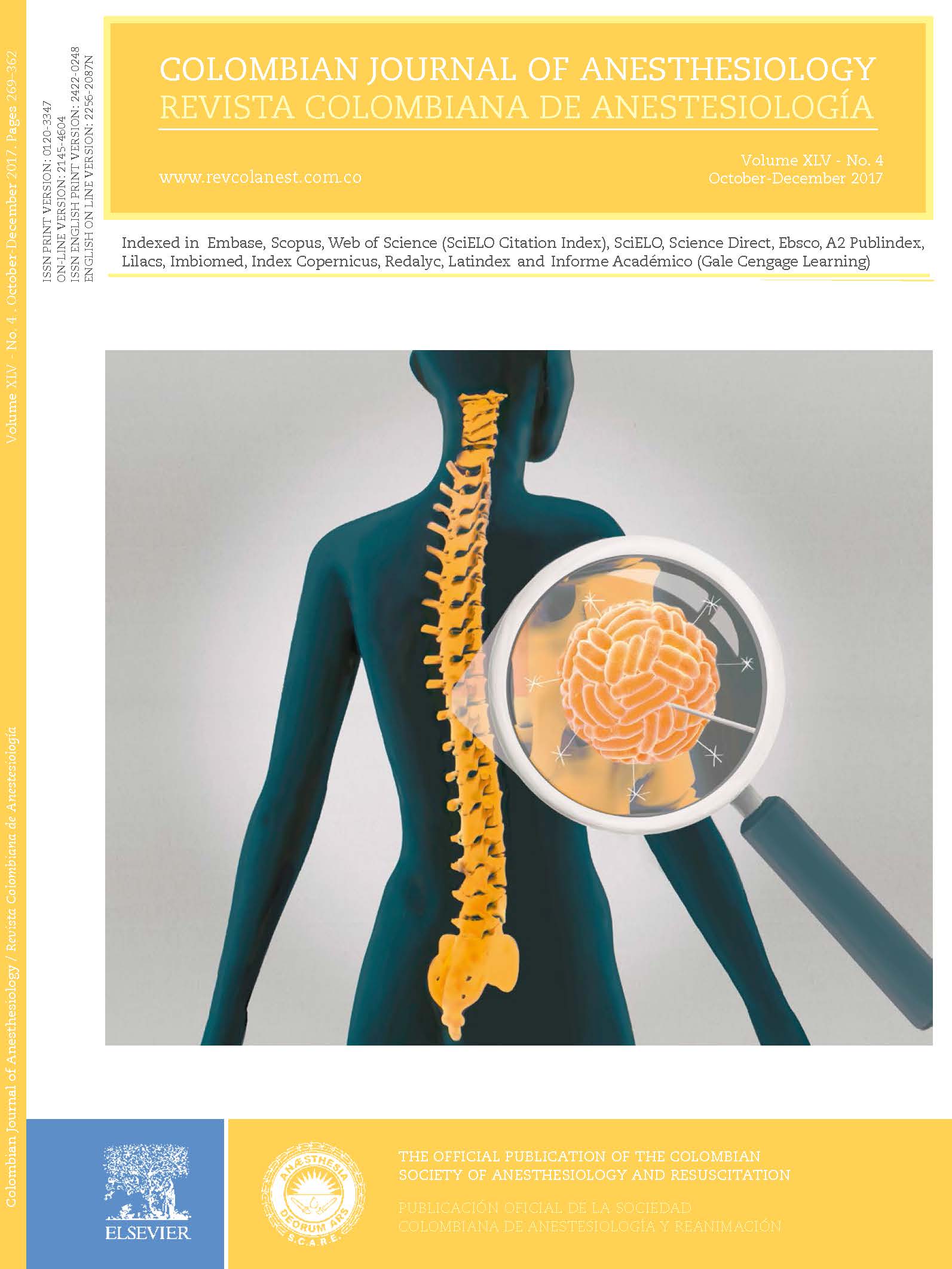
Ultrasound guided supraclavicular perivascular block. Anatomical, technical medial approach description and changes in regional perfusion
Abstract
Introduction:
Supraclavicular block is usually performed using a lateral to medial approach, although a medial to lateral approach is also feasible. Block onset may be evaluated through the sympathetic effect associated with the sensitive and motor blockade.
Objective:
To describe the ultrasound-guided supraclavicular block using a medial approach, evaluating the sensitive, motor, and sympathetic block onset.
Materials and methods:
An ultrasound-guided supraclavicular block was performed in a fresh cadaver with 20 ml volume (2 ml of iodine and 1 ml of methylene blue). A CT scan was performed and sagittal sections were obtained. The clinical phase included 10 patients undergoing a medial approach block; the onset of the block was evaluated based on a motor, sensory and sympathetic assessment (measuring flow changes in the humeral artery, the palmar temperature, and the perfusion index).
Results:
Adequate distribution of the contrast medium was observed in the cadaver, with complete spread through the brachial plexus, both in terms of the CT-reconstruction as in the anatomical cross sections. A significant change in all the sympathetic block parameters was observed 5 min after the bock: temperature (32.5 ± 1.8 °C to 33.4 ± 1.7 °C; p = 0.047), humeral arterial flow (105 ± 70ml/min to192 ± 97ml/min; p = 0.007), and thumb perfusion index (5 ± 3 to 10 ± 3%; p=0.002). The block was effective and uneventful in all patients.
Conclusions:
This supraclavicular approach achieves a homogeneous distribution throughout the brachial plexus, with high anesthetic efficacy. Regional changes secondary to the sympathetic block occur early after the block.
References
2. Franco CD. The subclavian perivascular block. Tech Reg Anesth Pain Manag. 1999;3:212-6.
3. Miller RD. Anestesia regional. In: Miller RD, editor. Anestesia. Octava Ed. Barcelona, España: Elsevier; 2015. p. 1405-40.
4. Hadzic A. Hadzic's peripheral nerve blocks and anatomy for ultrasound-guided regional anesthesia 2nd ed. New York: McGraw-Hill; 2012. p. 4216-33.
5. Kulempkaff D. Die Anasthesierung des plexus brachialis. Zentralbl Chir. 1911;38:1337.
6. Winnie AP, Collins VJ. The subclavian perivascular technique of brachial plexus anesthesia. Anesthesiology. 1964;25:353-63.
7. Brown DL, Cahill DR, Bridenbaugh LD. Supraclavicular nerve block: anatomic analysis of a method to prevent pneumothorax. Anesth Analg. 1993;76:530-4.
8. Pham-Dang C, Guns JP, Gouin F, Poirier P, Touchais S, Meunier JF, et al. A novel supraclavicular approach to brachial plexus block. Anesth Analg. 1997;85:111-6.
9. Perlas A, Lobo G, Lo N, Brull R, Chan VW, Karkhanis R. Ultrasound-guided supraclavicular block: outcome of510 consecutive cases. Reg Anesth Pain Med. 2009;34:171-6.
10. De Andrés J, Sala-Blanch X. Ultrasound in the practice of brachial plexus anesthesia. Reg Anesth Pain Med. 2002;27:77-89.
11. Subramanyam R, Vaishnav V, Chan VW, Brown-Shreves D, Brull R. Lateral versus medial needle approach for ultrasound-guided supraclavicular block: a randomized controlled trial. Reg Anesth Pain Med. 2011;36:387-92.
12. Marhofer P, Harrop-Griffiths W, Willschke H, Kirchmair L. Fifteen years of ultrasound guidance in regional anaesthesia: Part 2. Recent developments in block techniques. Br J Anaesth. 2010;104:673-83.
13. Soares LG, Brull R, Lai J, Chan VW. Eight ball, corner pocket: the optimal needle position for ultrasound-guided supraclavicular block. Reg Anesth Pain Med. 2007;32:94-5.
14. Chan VW, Perlas A, Rawson R, Odukoya O. Ultrasound-guided supraclavicular brachial plexus block. Anesth Analg. 2003;97:1514-7.
15. Feigl G1,Fuchs A, Gries M, Hogan QH, Weninger B, Rosmarin W. A supraomohyoidal plexus bloc designed to avoid complications. Surg Radiol Anat. 2006;28:403-8.
16. Galvin EM, Niehof S, Verbrugge SJ, Maissan I, Jahn A, Klein J, et al. Peripheral flow index is a reliable and early indicator of regional block success. Anesth Analg. 2006;103:239-43.
17. Iskandar H, Wakim N, Benard A, Manaud B, Ruel-Raymond J, Cochard G, et al. The effects of interscalene brachial plexus block on humeral arterial blood flow: a Doppler ultrasound study. Anesth Analg. 2005;101:279-81.
18. Minville V, Gendre A, Hirsch J, Silva S, Bourdet B, Barbero C, et al. The efficacy of skin temperature for block assessment after infraclavicular brachial plexus block. Anesth Analg. 2009;108:1034-6.
19. Hermanns H, Braun S, Werdehausen R, Werner A, Lipfert P, Stevens MF. Skin temperature after interscalene brachial plexus blockade. Reg Anesth Pain Med. 2007;32:481-7.
20. Winnie AP, Radonjic R, Akkineni SR, Durrani Z. Factors influencing distribution of local anesthetic injected into the brachial plexus sheath. Anesth Analg. 1979;58:225-34. [ Links ]
21. Tutoglu A, Boyaci A, Kücü A, Çakalar A, Sert H, Yalcm §. Perfusion index is increased in acute complex regional pain syndrome type 1. Arch Rheumatol. 2015;30:40-4.
22. Li J, Karmakar MK, Li X, Kwok WH, Ngan Kee WD. Regional hemodynamic changes after an axillary brachial plexus block: a pulsed-wave Doppler ultrasound study. Reg Anesth Pain Med. 2012;37:111-8.
23. Kus A, Gurkan Y, Gormus SK, Solak M, Toker K. Usefulness of perfusion index to detect the effect of brachial plexus block. J Clin Monit Comput. 2013;27:325-8.
24. Yang CW, Kwon HU, Cho CK, Jung SM, Kang PS, Park ES, et al. A comparison of infraclavicular and supraclavicular approaches to the brachial plexus using neurostimulation. Korean J Anesthesiol. 2010;58:260-6.
Downloads
Altmetric
| Article metrics | |
|---|---|
| Abstract views | |
| Galley vies | |
| PDF Views | |
| HTML views | |
| Other views | |


